FINLAND
Finnish Advanced Microscopy Node
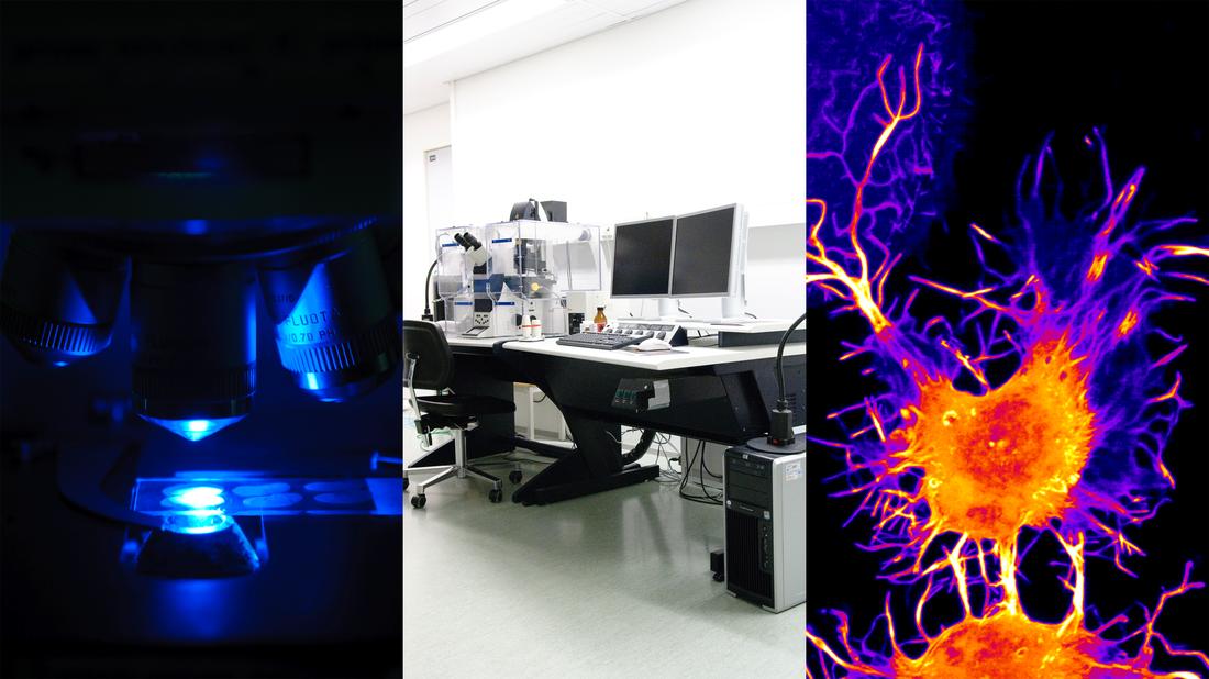
The Finnish Advanced Microscopy Node is composed of imaging facilities at three sites of Helsinki, Turku, and Oulu that have long experience in working together and offering open-access imaging technologies and expert services to both academic and industrial users. Annually, the Finnish Node serves more than 800 national and international users. The Finnish Euro-BioImaging Node has strong expertise especially in 3D services across multiple platforms in light and electron microscopy, and is capable of providing advanced 3D imaging, also in areas where this is not always commonplace. The Finnish Node offers its services as comprehensive 3D packages that include both 3D image acquisition, 3D reconstruction, and image analysis in 3D.
Read the news from the Finnish Advanced Microscopy Node
New offer of technology
The Finnish Node has started to offer a new imaging technology to Euro-BioImaging users. CARS (Coherent Anti-stokes Raman Scattering) microscopy is a label-free imaging technology that allows for chemically specific imaging of biological materials without the use of fluorescent labels. This technology is available in Helsinki using Leica TCS SP8 microscope (two HyD detectors, one PMT detector, resonant scanner, and full environmental chamber). Dedicated support is provided to Euro-BioImaging users by CARS microscopy expert. Our Leica SP8 system is equipped with a tunable dual-wavelength picosecond light source for multi-photon excitation. Apply now to conduct your imaging experiment using CARS microscopy at the Finnish Node.

Specialties and expertise of the Node
The Finnish Node focuses on four specialties that are state-of-the-art imaging technology areas: super-resolution imaging, correlative light and electron microscopy, mesoscopic imaging, and label-free technologies. Examples of research application areas that are well covered in Finland with these technologies are metabolism and inflammation, developmental biology, cell migration, adhesion and invasion, quantitative cell biology, extracellular matrix biology, organotypic cultures, cardiovascular research, neurology, and cancer biology. All of these research applications cover a wide spectrum of model organisms and in vitro models. Bioimage bioinformatics and image analysis are another area of expertise of the Finnish Euro-BioImaging Node and all three Node partners composing the Finnish Node engage in significant software development. The Finnish Node offers image analysis platforms such as BioimageXD, an open-source platform for processing, visualization and analysis of multidimensional image data and Microscopy Image Browser (MIB), freely available software for image processing, segmentation, and visualization of multidimensional (2D-4D) light and electron microscopy data. In addition, the Centre for Machine Vision and Signal Analysis Research has extensive expertise in computer vision assisting Euro-BioImaging users in Oulu with their image analysis needs.
Technologies:
| Offered Technologies |
| Deconvolution Widefield Microscopy (DWM) |
| Laser scanning confocal microscopy (LSCM/CLSM) |
| Immuno-electron microscopy (immunogold labeling of Tokuyasu cryosections) |
| 3D Scanning Electron Microscopy (SEM) of plastic embedded samples |
| Image Scanning Microscopy (ISM) |
| Array Tomography |
| Serial Block Face Scanning EM (SBF-SEM or SBEM) |
| Serial section Transmission Electron Microscopy (ssTEM) |
| TEM Tomography |
| Spinning disc confocal microscopy (SDCM) |
| Total internal reflection fluorescence microscopy (TIRF) |
| Stimulated emission depletion microscopy (STED) |
| Focused Ion Beam SEM (FIB-SEM) |
| Genetic encoded EM probes (GE probes) |
| Large scale EM/TEM (IsEM/lsTEM) |
| pre-embed Correlative Light and Electron Microscopy (pre-embed CLEM) |
| pre-embedding Immunolabelling (pre-embed IL) |
| Immuno-gold EM on resin sections (resin-EM) |
| Second/Third Harmonic Generation (SHG/THG) |
| Single Molecule Localization Microscopy (SMLM) |
| Stochastic optical reconstruction microscopy (STORM) |
| CLEM from live cells to plastic sections ("live" CLEM) |
| Fluorescence correlation spectroscopy (FCS) |
| Fluorescence cross-correlation spectroscopy (FCCS) |
| Fluorescence-lifetime imaging microscopy (FLIM) |
| Fluorescence resonance energy transfer (FRET) |
| Fluorescence recovery after photobleaching (FRAP) |
| Optical projection tomography (OPT) |
| Selective plane illumination microscopy (SPIM) |
| Multiphoton microscopy systems |
| Photo activated localization microscopy (PALM) |
| Coherent Anti-stokes Raman Scattering (CARS) |
| Atomic Force Microscopy (AFM) |
| Traction Force Microscopy (TFM) |
| Image Analysis-bio (IA-bio) |
| Mass Spectrometry-based Imaging-bio (MSI-bio) |
| High-throughput Microscopy (HTM) |
Additional services offered by the Node
- Methodological setup (e.g. design of study protocols and standard operation procedures)
- Technical assistance to run instruments
- Animal facilities
- Wet lab space
- Data processing and analysis
- Training seminar rooms
- Cell culture facilities - Safety Levels 1 and 2

Instrument highlights
Special features of instruments at the Finnish AM Node are:
- The combination of different imaging techniques such as correlative light and electron microscopy (CLEM) that combines electron microscopy with transmitted light or fluorescence microscopy.
- The use of 3D imaging in super-resolution imaging techniques such as 3D tomographic STED (mirror-STED) and 3D in super-resolution localization techniques with N-STORM.
- Mesoscopic-scale multidimensional (3D-4D) imaging using optical projection tomography (OPT) and light sheet fluorescence microscopy (LSFM)/ selective plane illumination microscopy (SPIM).
Testimonials
“The cell imaging core facility at Turku University is very well managed with dedicated professional people. The instruments are of high quality and are very well maintained. I was very pleased to be here and I am satisfied by the experience of coming here for a few days and having the imaging done perfectly.”
- Jean-Claude Twizere, IR, Ph.D. - University of Liège
“The visit to the Euro-Bioimaging Finnish Node changed the direction of my research in a positive way: I was able to observe the behaviour of my protein of interest in really high resolution using SIR microscopy. The staff were always ready to help and guide me to get the best out of my visit. I highly recommend a visit to the Finnish Node!”
- Paulina Moreno Layseca Ph.D - Universitätsklinikum Hamburg-Eppendorf
Contact details
Irina Belaia
Finnish Advanced Microscopy Node Manager
Contact-FiALM@eurobioimaging.fi
www.eurobioimaging.fi