UNITED KINGDOM
The UK Node
The UK Node offers open access to a wide range of advanced biological imaging techniques, including correlative, multi-modal, high-content and super-resolution imaging. It is a multi-sited national infrastructure hosted by twelve leading institutions across the UK (see below for site details). Together, they offer state-of-the-art imaging equipment, expertise, training and image data services. In addition, the UK Node includes a distributed sub-Node, UK-BIAS (UK National Core Facility for Bioimage Analysis), bringing the total to thirteen sites across the UK.
These technologies can be applied to a wide range of fundamental and translational research projects at molecular to cellular resolution — from single cells to 3D in vitro models and whole organisms.
The Euro-BioImaging UK Node Manager acts as the single point of contact who can direct potential users to the relevant imaging technology and site to undertake their experiments. We welcome applications from all research disciplines. Support will be provided for the entire experimental pipeline including (where appropriate) initial user consultation, experimental planning, full hands-on training, assistance with sample preparation, gathering highest quality imaging data and data analysis.
FUNDING IS AVAILABLE: UK researchers can apply for up to £5K for hardware access and image analysis or up to £2K for image analysis services only – please see here for more details.
Read the news from the UK Node
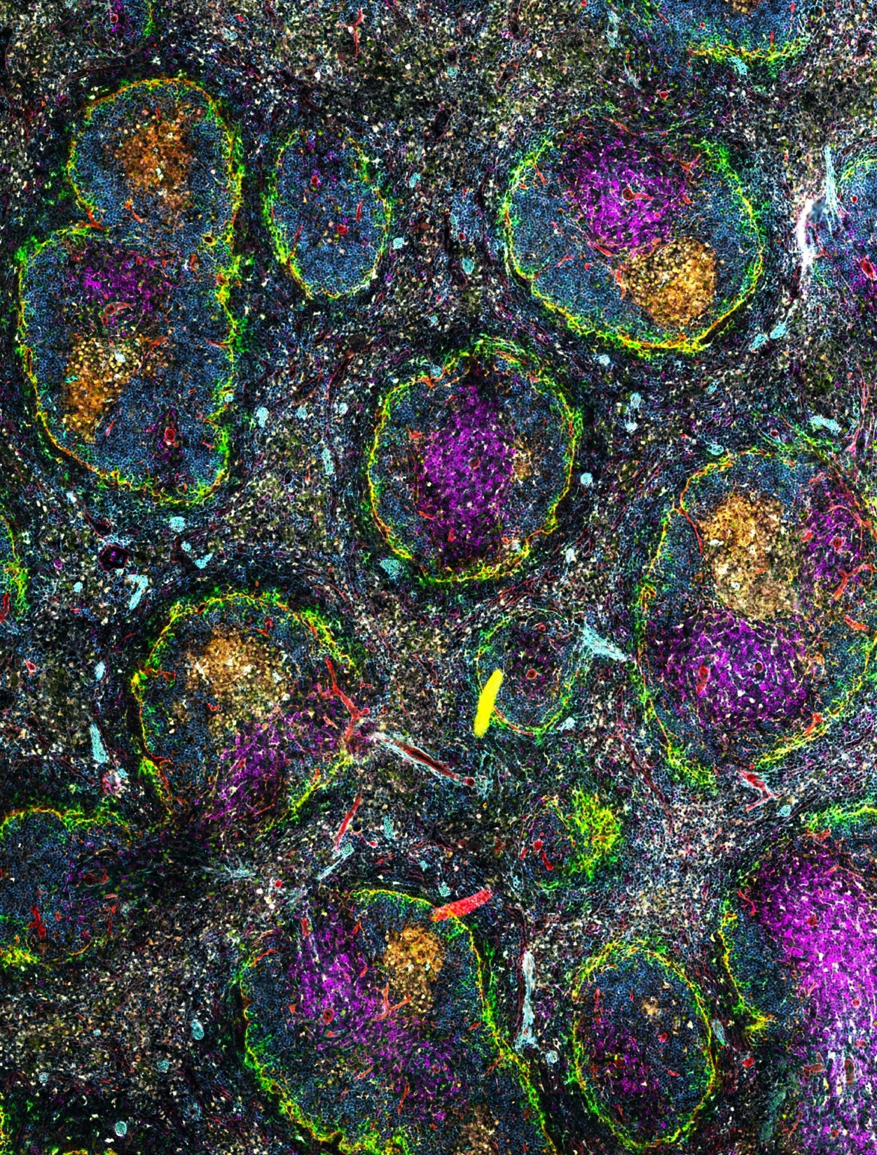 Spatial proteomics using the MACSima system on a mouse germinal centre acquired by Isabel San Martin Molina and Edith Marcial Juarez at the Babraham Institute.
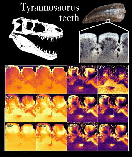 LA-ICP-MS image of Tyrannosaurus teeth acquired by Dr Aaron LeBlanc, KCL.
Specialities and Expertise of the UK Node
Babraham Institute:
The Babraham Institute Imaging Facility provides access to advanced imaging technologies for light and electron microscopy. We offer a Molecular Devices HT.ai with robotic plate loader for high throughput/high content imaging, a Zeiss Axioscan 7 for high-throughput slide scanning, a Miltenyi UltraScope Blaze for mesoscopic light-sheet imaging and a Miltenyi MACSima for spatial proteomics. For electron microscopy the Facility has a Zeiss CrossBeam 550 FIB SEM enabling standard SEM/STEM imaging, and volumetric EM using either ion beam milling or array tomography. With technologies available for both light and electron microscopy, the Facility fully supports correlative imaging experiments, including 3D volumetric data acquisition, alignment and quantification. Experienced staff will guide, train and assist users across the complete workflow from sample preparation to image acquisition, image processing and image analysis.
University of Birmingham:
The University of Birmingham Advanced Imaging Facilities provide open access to a highly integrated, multi-modal bioimaging environment spanning fluorescence microscopy from single molecules to cells, tissues and whole organisms. The Facilities offer expertise in ultrafast single-molecule and functional imaging, super-resolution microscopy, light-sheet and deep-tissue imaging, multiphoton microscopy, advanced sample preparation and quantitative image analysis. Users are supported across the full experimental workflow, from project design, sample preparation and training through to data acquisition, analysis and interpretation. Embedded within Birmingham’s interdisciplinary research environment, including the Centre of Membrane Proteins and Receptors (COMPARE), clinical research infrastructure and computational expertise, the Facilities provide a collaborative platform where advanced microscopy, probe development, custom imaging technologies and quantitative analysis can be applied to biological and biomedical discovery.
ESRIC:
The Edinburgh Super-Resolution Imaging Consortium (ESRIC) comprises open access advanced imaging facilities specialising in super-resolution techniques. As well as cutting-edge equipment, we bring together a broad spectrum of knowledge and expertise in extending the boundaries of biological imaging beyond the diffraction limit. Current technologies include dSTORM/PALM/DNA-PAINT on Nikon N-STORM and Olympus Cell Excellence systems, gSTED, Tau STED and FLIM-FRET on a Leica SP8 with FALCON FLIM, Structured Illumination Microscopy on a Nikon N-SIM /SoRa hybrid system, and SRRF on an Andor Dragonfly spinning disk. Our equipment enables us to image objects in 3D at super-resolution in up to 4 colours, with resolutions ranging from 120nm down to 10nm.
Francis Crick Institute:
Our Light-sheet systems cover two distinct application spaces: larger fixed samples (1 - 20 mm) can be imaged with the Miltenyi-LaVision Ultramicroscope II whereas smaller living or fixed samples (50 - 1000 µm) can be imaged with the Leica Viventis Deep. The Ultramicroscope can be used in combination with antibody labelling and tissue clearing to image many sample types, including whole mouse organs, later-stage embryos, tumours, and tissue sections. In contrast the Leica Viventis Deep, thanks to the dual detection, is suitable for deep live and fixed imaging of small samples such as spheroids, organoids and early-stage embryos with a medium throughput.
HREM is a block-face imaging technique for the analysis of tissue and organ morphology that does not require endogenous tissue fluorescence or antibody labelling but is compatible with lacZ and NBT/BCIP staining. It has been widely used to study cardiovascular, skeletal, and neurological defects in mouse models (e.g. Vanyai et al., Development, 2020)
King’s College London:
At the atomic and nano scales, King's Centre for Electron Microscopy (KCEM) supports advanced imaging techniques, including Transmission Electron Microscopy (TEM), Scanning Electron Microscopy (SEM), Serial Block Face SEM (SBF-SEM), Correlative Light and Electron Microscopy (CLEM), TEM Tomography, Scanning Transmission Electron Microscopy (STEM), and Energy Dispersive X-ray Spectroscopy (EDS), serving researchers across medicine, biological sciences, materials science, and engineering.
Moving down the resolution scale, we provide access and support in, super-resolution and low-toxicity live cell imaging (N-STORM 5.0 and SoRa, plus single and multi-photon lightsheet capability), as well as high-content and throughput spinning disc (Opera Phenix).
Extending beyond optical imaging, we deliver analytical multi-modal imaging in the form of 2D or 3D spatial quantitative elemental and molecular imaging of both endogenous elements and metal tagged antibodies in cells and tissues using a multiplexing methodology whilst identifying associated lipids and biomolecules in a label free approach. Routine LA-ICP-MS (Laser Ablation Inductively Coupled Plasma Mass Spectrometry) has been integrated with DESI (Desorption electrospray ionisation)-MS and Raman Spectroscopy as a single unique correlative workflow. Together with integration of IR, this workflow will enable high resolution spatial mapping of lipid, nucleic acids and protein phosphorylation.
University of Leeds:
The Leeds Node integrates four complementary facilities: the Bioimaging Facility (STED, SMLM, Lattice Lightsheet, Airyscan), the CryoEM Facility (two Titan Krios microscopes and a Hydra Bio FIB-SEM with in-chamber fluorescence for CLEM), the UK-unique HyBiFa hyperspectral platform enabling simultaneous label-free chemical, structural and functional imaging via SRS, CARS, SHG and FLIM, and an AFM Facility housing nine atomic force microscopes including ultra-high-speed biological imaging systems and optical tweezers. Together these platforms support a globally leading multi-scale imaging pipeline spanning live-cell imaging through to near-atomic resolution structural biology.
University of Liverpool:
At Liverpool, we provide an integrated ensemble of imaging modalities in one place for imaging across scales from single molecule to cells and model organisms. Example projects include cell biology, surface science, biochemistry, and microbiology. Of particular note is our unique Bio AFM-TIRF imaging system for atomic force microscopy combined with dual fluorescence total internal reflection. We support the entire experimental workflow for each technique with dedicated staff, and we use an OMERO server for data management.
Newcastle University:
Newcastle University provides a comprehensive spatial biology site that integrates world-class imaging technologies, tissue resources, genomics, bioinformatics and data science into a single end-to-end integrated workflow. Through access to extensive tissue collections, including archived and freshly collected clinical samples, researchers can seamlessly progress from tissue acquisition and processing through effective labelling strategies to advanced spatial analysis, enabling the study of gene and protein expression within their native biological context. Our capabilities are underpinned by established research facilities spanning, histology, bioimaging, genomics, bioinformatics and human tissue resources (biobanks), supported by expertise in tissue labelling, imaging, spatial transcriptomics and multimodal data integration.
An example is the SpaRTAN (Spatially Resolved Transcriptomics at Newcastle) service, which is manufacturer-certified and has operated since 2020. It delivers a complete spatial biology workflow, from experimental design and tissue preparation through sequencing, spatial imaging and advanced bioinformatic analysis. SpaRTAN brings together expertise from Newcastle’s Bioimaging Facility, Genomics Core Facility, Bioinformatics Support Unit and Human Developmental Biology Resource, providing researchers with a coordinated route from sample to biological insight.
By combining molecular, cellular and tissue-scale information, our site enables researchers to move beyond traditional single-modality approaches and uncover the spatial relationships that drive health, development and disease, accelerating discovery and translational impact across the biomedical sciences.
CLF OCTOPUS:
We offer multiple microscopy techniques on a suite of commercial and custom-built systems, including MINFLUX, light sheet, single molecule localisation microscopy (PALM/STORM, including cryo-STORM), STED, single molecule methods including tracking and Fluorophore Localisation Imaging with Photobleaching (5 nm precision), confocal (including two photon and FRET/FLIM), optical trapping, and cryo focused ion beam SEM.
The microscopes are co-located with extensive preparation facilities including laboratories for biochemistry, cell biology, and chemistry. Facility users have access to a dedicated support team including microscopists, cell and molecular biologists, chemists, and data analysis experts. Users are provided with assistance at all stages of the experiment, including sample preparation and labelling, collection of data, and data analysis and interpretation. Custom data analysis packages are available and can be tailored to the requirements of a particular experiment. Accommodation and catering facilities are available on campus.
University of Oxford:
Oxford's Advanced Microscopy Core brings together the Micron Bioimaging Facility, the Cellular Imaging Core Facility (CICF) and the Oxford-Zeiss Centre of Excellence in Biomedical Imaging. Oxford leads nationally in super-resolution, offering 3D-SIM, TIRF-SIM, iSIM, Lattice SIM, TauSTED and SMLM alongside staff-developed quality-control software (SIMCheck, SIMWorks) for artefact-free imaging. The Oxford-Zeiss Centre adds Lattice Light-Sheet and functional imaging (FCS, FCCS, FLIM), while CICF's CL3-enabled facilities support high-content, organoid and iPSC workflows. Integrated analysis infrastructure spanning Arivis, Imaris, Aivia and ML/deep-learning pipelines supports complex 3D/4D datasets, and the Core meets international QUAREP-LiMi quality standards.
Oxford Brookes University:
We focus on providing volumeEM and CLEM workflows, including dual axis serial section cellular electron tomography and serial block face-scanning electron microscopy (SBF-SEM). We can work with fixed or high pressure frozen samples and have extensive expertise in eukaryotic parasite work and insect vectors. The centre is equipped with CATII culturing facilities within the Centre, as well as a SAPO licence to support these projects.
UK-BIAS:
UK-BIAS (UK National Core Facility for Bioimage Analysis) is a distributed sub-Node of expert bioimage analysts based across ten UK institutions, providing FAIR-compliant, reproducible image analysis pipelines. The team has extensive experience with open-source platforms widely used by the biosciences community — including ImageJ/FIJI, CellProfiler, ilastik, QuPath and napari — as well as developing bespoke analysis pipelines in Python, Java and MATLAB. They support projects from initial consultation through to long-term collaboration, with all pipelines released as open-source software.
University of York:
Current technologies include PALM/STORM/SIM2 via the Zeiss ELYRA 7, Spatial-omics via Nanostring GeoMX-DSP and 10X Visium HD, Label-free imaging via PhaseFocus LiveCyte and Tomocube and multiphoton imaging via Zeiss LSM980 AiryScan2 along with the more routine confocal, slide scanning and volume (serial blockface and array tomography) electron microscopy with elemental analysis, imaging flow cytometry (BD FACSDiscover S8) and Mass Spec Imaging. The team have a wide breadth of biological expertise spanning plant sciences through to biomedicine, and are familiar with most sample types and sample preparation methods, including materials science.
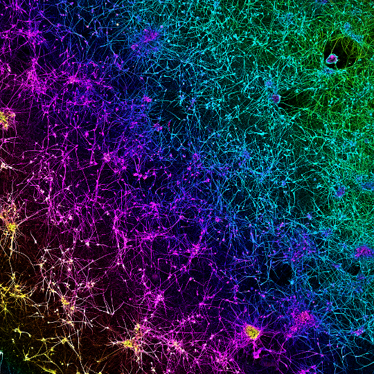 Dopamine network acquired by Nick Gatford, at the Micron Facility, part of University of Oxford.
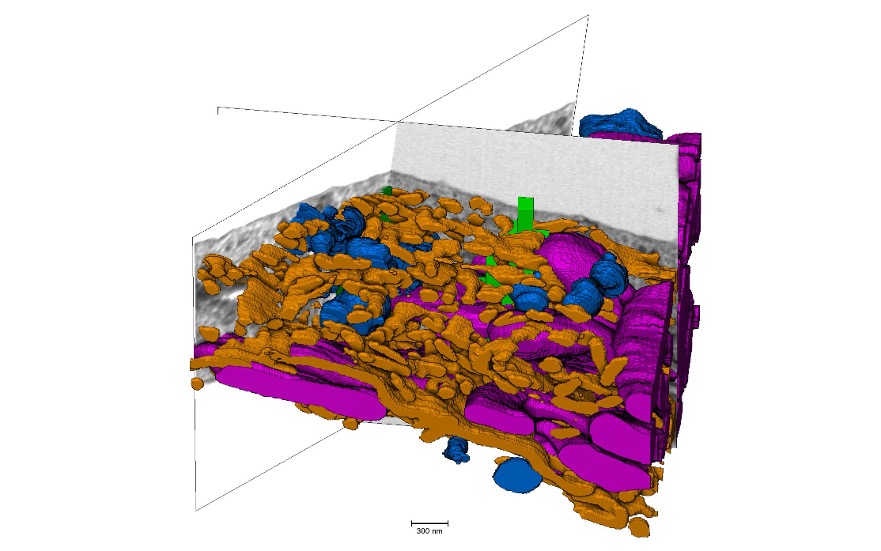 vEM dataset from a HEK cell acquired by Kirsty MacLellan-Gibson, Nick Ktistakis and Maria Manifava at the Babraham Institute.
Additional services offered by the Node
- Project planning and management
- Wet lab
- Cell culture facilities
- Methodological setup
- Facility induction
- Technical assistance to prepare experiments and run instruments
- Workstations
- Data storage
- Image acquisition
- Image processing and analysis
Probes
All sites within the EuBI UK Node operate within leading edge academic institutions and as such, many different types of probes, including plasmids, antibodies and chemical reagents are available within individual researcher labs. Additionally, a number of facilities maintain their own in-house reagents, including pre-labelled antibodies for super-resolution (STORM) and spatial transcriptomics.
Other model organisms
All sites within the EuBI UK Node operate within leading edge academic institutions that host labs working with a broad range of different model organisms, including zebrafish, Drosophila and c.elegans. Facilities to support users working with such organisms would therefore be possible, subject to discussions and agreements with the appropriate academics.
High biological safety level
BSL2 facilities are available within a number of the laboratories at the host institutions for the EuBI UK Node facilities and can be made available to users following consultation with the site leads. Of note, Oxford Brookes holds a SAPO license for culturing Trypanosoma brucei within the imaging facility itself. BSL3 facilities are also available at Newcastle University and University of Oxford where both sites have extensive experience supporting users with BSL3 infectious material or pathogens.
Contact details
Georgina Fletcher
EuBI UK Node Manager
georgina@rms.org.uk