MagnetoEncephaloGraphy (MEG) *
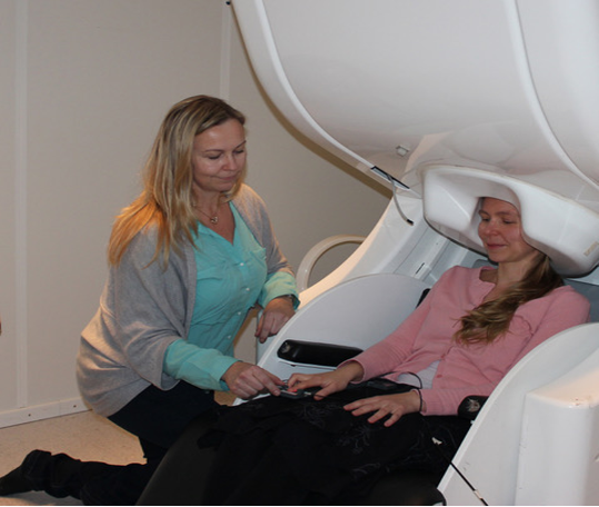
Magnetoencephalography (MEG) measures noninvasively minute magnetic fields produced by neuronal activity of the brain. In electroencephalography (EEG), a voltage distribution produced by neuronal currents is measured with scalp electrodes whereas in MEG neuronal activity is detected by superconducting sensors measuring the corresponding magnetic fields outside the head.
Source modelling is used in MEG to model, quantify, and localize sources contributing to the measured magnetic fields at ms temporal resolution and a few mm spatial resolution. Skull, cerebrospinal fluid, and scalp are transparent to magnetic fields. MEG is optimally suited for detecting neuronal dynamics in the cerebral cortex. The first whole-head commercial MEG systems were introduced in the early 90’s. At present, MEG systems are widely used in hospitals and academic research infrastructures.
The potential clinical and research uses include for example studies of the functions of neural oscillations, the nature of event-related brain activation, mechanisms of functional connectivity between regions and the emergence of modes of network communication in brain systems.
MEG has two clinical applications related to the localizations of epileptic foci and mapping functionally important regions in the brain prior to surgery.
REFERENCES.
S. Baillet, “Magnetoencephalography for brain electrophysiology and imaging”, https://doi.org/10.1038/nn.4504
J. Gross, “Magnetoencephalography in Cognitive Neuroscience: A Primer”, https://doi.org/10.1016/j.neuron.2019.07.001
R. Haari et al., “IFCN-endorsed practical guidelines for clinical magnetoencephalography (MEG)”, https://doi.org/10.1016/j.clinph.2018.03.042
MEG is available at the Finnish Biomedical Imaging Node.
MEG Use cases:
| Use cases | Node | DOI |
|---|---|---|
| Assessment of the developmental vs progressive character of the impairment of spinocortical proprioceptive pathways in Friedreich ataxia | Finnish Biomedical Imaging Node | https://doi.org/10.1212/wnl.0000000000007750 |
| Study of the neural underpinnings of predictive processing in natural speech | Finnish Biomedical Imaging Node | https://doi.org/10.1016/j.neuroimage.2020.116936 |