Micro X-ray Fluorescence Spectrometry (XRF)
Micro-X-Ray Fluorescence Spectrometry (XRF) allows the qualitative analysis of the elemental composition of solid samples. The detected signal results from the emission of element-characteristic fluorescence X-rays from a sample bombarded with high-energy X-rays. The result is a spatially resolved distribution of elements represented in a 2D map. Possible applications include the investigation of element distribution in tissues, like bone, uptake of specific elements by tumorous tissue or foreign bodies within tissues and organs, e.g. implants.
Different XRF setups are available, which offer confocal (3D) or non-confocal (2D) modes. One setup uses polychromatic radiation from a Rhodium (20W) X-ray tube for excitation of low-, medium- and high-Z elements. Measurements can be performed in air or vacuum. The fluorescence signal is detected using a Si(Li) detector with an ultrathin window, which extends the detectable elements range from Uranium down to Magnesium or Sodium if in vacuo. Scans can be performed with spatial resolution of about 50 µm. A second micro-XRF setup uses focused monochromatic Mo-Ka radiation from a 2 kW Mo anode X-ray tube. The polycapillary optics produces a beam of 15 µm diameter. Measurements can be performed only under ambient conditions, so light elements are not accessible. The monochromatic excitation leads to lower detection limits than polychromatic excitation.
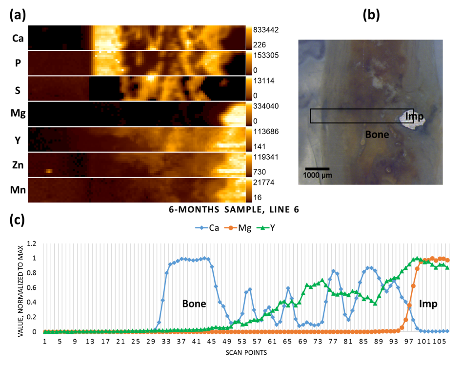
 Element distributions in bone carrying bioresorbable Mg implant.
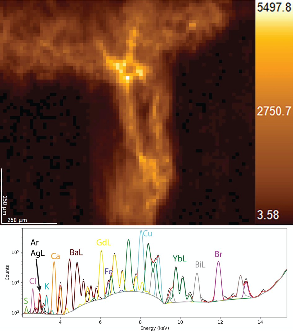
 2D Gd-Lα map from a gallbladder stone. The scan was conducted with a step size of 15
μm and a counting time of 50 s/point.