An open source image analysis tool for studying neuroinflammation linked to COVID-19
Published: 2023-11-15
COVID-19 infection has been associated with many neurological manifestations, from neuroinflammation to impaired brain energy metabolism. In the Turku PET Centre, which is part of Euro-BioImaging’s Finnish Biomedical Imaging Node, autoradiography (ARG) is used to study brain inflammation linked to COVID-19. While ARG provides high resolution images of brain tissue, analysis of the obtained data can be very laborious. With support from the ISIDORe project, an open source, user-friendly, automated analysis pipeline was developed for aligning and processing ARG images from mouse brain. This so-called Mouse Brain Alignment Tool (MBAT) helps to increase efficiency, accuracy and reproducibility in image registration and analysis in studies aiming to better understand the characteristics and mechanisms of neuroinflammation. We spoke to the developers - Zuzana Čočková, a neuroscientist working at Charles University, Prague, and Junel Solis, an image analyst at Turku BioImaging, part of our Finnish Advanced Microscopy Node - to learn more about this tool and their work.
Zuzana Čočková is a neuroscientist working at Charles University. During her PhD, she traveled to Turku, Finland, for an internship at the renowned Turku PET Centre, in the team of Francisco López-Picón. While she was there, her mission was to better understand neuroinflammation linked to COVID-19 infection.
In brain research, various tracers are commonly used to explore different aspects of brain health and dysfunction. Indeed, recent articles have utilized inflammation tracers (e.g., FDPA) or metabolic tracers (e.g., FDG) together with CNS PET imaging, revealing profound inflammation in the brains of long COVID-19 patients (DOI: 10.1007/s00259-021-05215-4, 10.1101/2022.06.02.22275916).
In the Turku PET Centre, autoradiography (ARG) is used to study brain function, because the resolution is higher compared to PET. This method aims to visualize the anatomical distribution of a protein of interest in tissue from experimental animals with the ultimate goal being quantification of photostimulated intensity per unit in brain regions of interest.
Analyze specific regions of brain inflammation
“Depending on the specific marker used, you can investigate brain inflammation or measure the degeneration of synapses,” explains Zuzana. “You choose a marker and you apply it. You make slices of the brain, then you look at specific regions. The inflammation is not happening on the same scale everywhere.”
However, the image analysis with ARG is quite laborious. Individual regions of interest are drawn manually every time, which has several drawbacks. “It’s time-consuming, and it’s not very objective,” explains Zuzana, who was involved with image analysis in her internship. “Everyone does it differently.”
It was over a lunchtime discussion with Joanna Pylvänäinen, image data analyst, then at Turku BioImaging, that the idea to develop an automated ARG image analysis workflow came about.
“Indeed, an automated workflow would be more robust. It would help us to do our job faster with more reliable results.”
ISIDORe funding to support the project
The problem is that Zuzana’s internship was nearly over. Enter ISIDORe funding to support infectious disease research. The Finnish Advanced Biological Imaging Node of Euro-BioImaging, to which Turku BioImaging contributes, was involved in the ISIDORe project through Euro-BioImaging. In addition, the Node provides image analysis as a stand-alone service through Euro-BioImaging, and is able to develop both biological and biomedical image analysis pipelines and tools for users, even if they acquire their images in a different facility.
Given the importance of her project to COVID-19 research, Zuzana applied for support from the ISIDORe project, a large-scale EU-funded project providing free transnational access to a portfolio of cutting-edge research services for studies on epidemic-prone disease. With ISIDORe funding, she returned to Finland, this time with the mission of developing the Mouse Brain Atlas Tool. Junel Solis, image analysis expert at Turku BioImaging, was tasked with developing the workflow in collaboration with Zuzana.
“The tool we developed with Zuzana is a Python-based software for processing autoradiographic (ARG) images of mouse brain tissue sections and analyzing them using topographical data from the Allen Institute’s Mouse Brain Atlas,” says Junel. “It automates the ARG image analysis process and makes it possible to focus on specific sub-regions of the brain,” explains Junel. “Automating the workflow was one aspect of this job. But we also paid special attention to ensuring that the workflow is user-friendly. It has to be packaged so people can download it easily, without administrator access.”
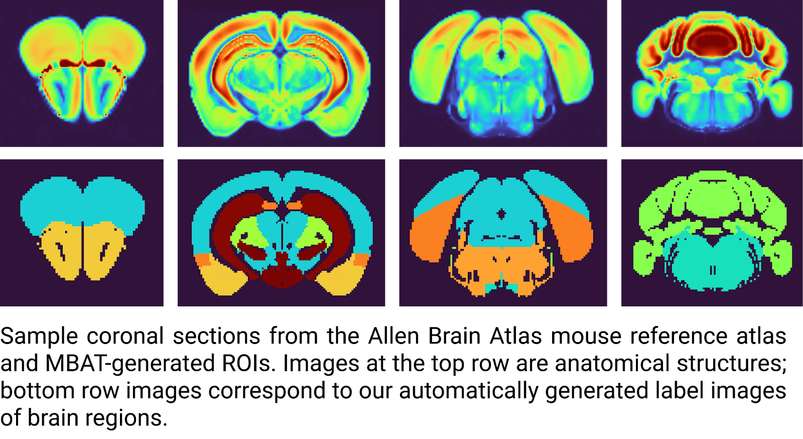
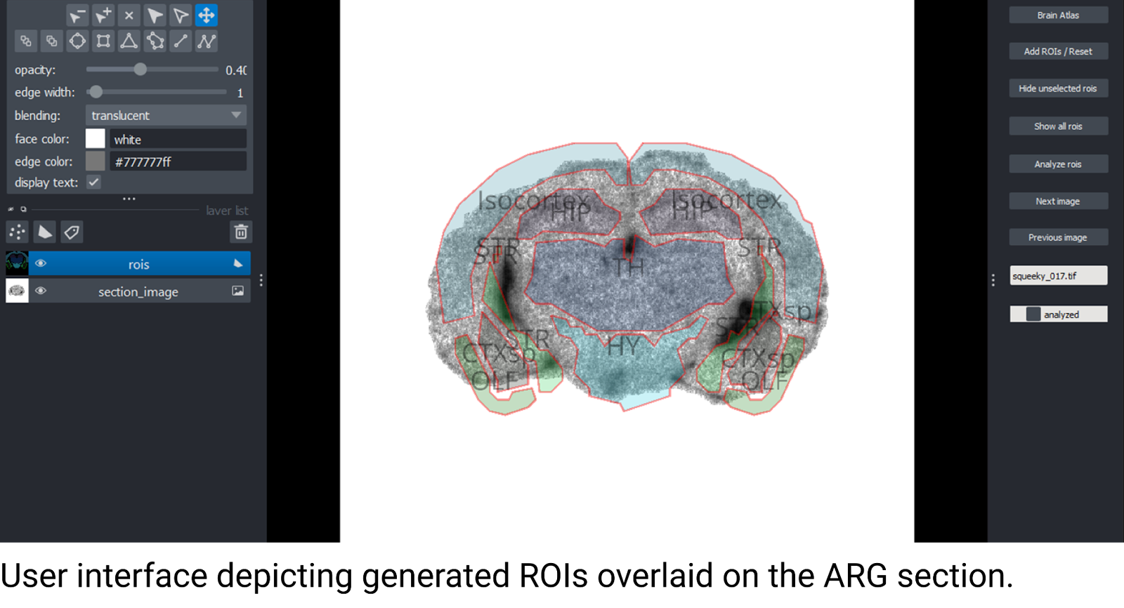
MBAT allows the researcher to preprocess their ARG sections, with different layout schemes contained in whole-slide images (WSI) and automatically convert those sections into single images. The included user interface allows the user to match brain sections with the atlas, and calculate mean background intensity. MBAT converts data from the Allen Institute’s Mouse Brain Atlas and stores the topographical information of various brain regions in a local database as regions-of-interest (ROI), which can then be overlaid onto the ARG image. The user can translate, rotate, scale, and even edit brain region ROIs. The ARG signal for each ROI can be measured and saved into a data file, which allows the user to process sections in smaller batches and return to the workflow at a later time.
Open science
Another criteria was that the software they develop should be open source. The Mouse Brain Alignment Tool developed by Zuzana and Junel is already on GitHub, an open access repository for sharing software tools and code, and ready to share. As the code is open, it becomes accessible to anyone, empowering them to make modifications according to their preferences.
“Without the ISIDORe funding, we wouldn’t have been able to develop this robust workflow and make it available in open access for the entire community,” explains Zuzana. “So we are extremely thankful that this project makes it possible to share these developments with a wider community.”
For example, this ISIDORe user project result could find its way to the BY-COVID project / registries, e.g. the Infectious Diseases Toolkit.
“The support of Zuzana’s Image Analysis user project at the Euro-BioImaging FiAM Node supported through the ISIDORe TNA Calls is also one great example for demonstrating the user-driven capacities behind the ISIDORe project and Euro-BioImaging ERIC operations. The overarching aim is to respond to researcher’s acute needs and provide access to the most suitable expertise and scientific services for addressing the research goals in a most efficient and timely way - in the overall mission to facilitate and advance research on the European landscape and beyond,” remarks Euro-BioImaging scientific project manager for ISIDORe, Arina Rybina.
How will this tool help other researchers/image analysts?
“Our tool is mainly focused on brain analysis, since a standardized brain atlas is applied,” explains Zuzana. “So in its current form, it is only applicable to the mouse brain. Adding more standardized brain atlases, for example for rat animal models is possible. And it was designed to work with ARG scans, so it would require some tuning to work with different types of image files. But this isn’t a big issue. In general, it should be useful for any application that aims for signal measurements in coronal slices of the brain, whether autoradiography or alternative staining methods, including fluorescent staining, were employed.”
“This tool could also be interesting for people working on brain autoradiography images set up on whole slides. In this case, it would require only small changes to make it applicable.”
We really encourage other researchers to check out the results of this ISIDORe-funded user project!
Get the Mouse Brain Atlas Tool on GitHub: https://github.com/turku-bioimaging/mouse-brain-alignment-tool